|
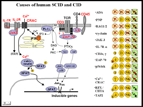
Figure A: Signal transduction in T cells: mutations
identified in severe combined immunodeficiency (SCID) and combined
immunodeficiency (CID) patients.

(left) The diagram depicts molecules involved in
signal transduction in T lymphocytes. Mutant proteins
implicated in the pathogenesis of human SCID are highlighted
in red. Jak3, janus kinase 3; IL-2Rg,
interleukin 2 receptor g chain (common
g chain, cg);
CRAC, calcium release activated calcium channel; ZAP-70, zeta-associated
protein 70; NFAT, nuclear factor of activated T cells; PLCg, phospholipase C g;
ER, endoplasmic reticulum.
(right) The presence or absence of T, B or NK cells in the
various forms of SCID and CID is indicated. Where the symbol
is crossed out, no cells are detectable. Low total T cell numbers,
low CD4+ and low CD8+ cell numbers, respectively, are indicated
by small circles. The arrow in the green box symbolizes T cell
activation; were crossed out, T cell activation is partially
(light red) or severely (dark red) impaired. E.g. patients with
a mutation in ZAP-70 have low to undetectable numbers of CD8+
T cells but CD4+, B and NK cell counts are normal. However, activation
of CD4+ cells is severely compromised. |
