|
Figure B: Partial, but unspecific, reconstitution of Ca2+ influx in the
presence of high extracellular Ca2+ and ionomycin concentrations.
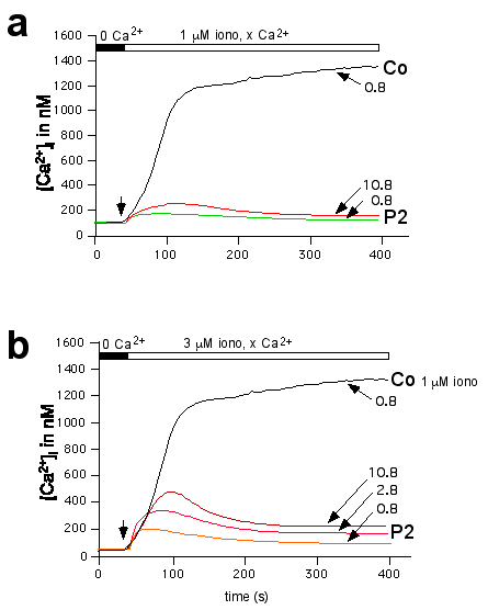
(a) Fura-2 loaded T cell lines from one SCID patient (P2)
and a normal control (Co) were stimulated with 1 mM
ionomycin in the presence of low (0.8 mM) or high (10.8 mM) extracellular
Ca2+. (b) T cells were stimulated with 3mM
ionomycin in the presence of 0.8 mM (control) and 0.8, 2.8 or
10.8 mM (patient 2) extracellular Ca2+, respectively. Under these
conditions, maximally attainable elevations in [Ca2+]i in the
SCID patients' T cells were transient and reached ~40% of normal
peak concentrations. This increase of [Ca2+]i in the patients' cells is most likely due to
ionomycin-mediated Ca2+ transport into the cell which is independent
of Ca2+ channels as the level of peak influx at 10mM [Ca2+]ex
corresponds to the ionomycin concentration (1x10⁻⁶ M and 3x10⁻⁶ M in
panel a and b, respectively).
|